
Login

Feb 9 – 12
We are hosting a live Myofascial Class with Pam West this weekend, so we will pick up things next week. In the meantime, we will leave you with more material to practice dry brush and Lymphatic Anatomy!
The dry brushing sequence is great for 2 reasons:
1) It is a very effective treatment if you are limited for time
2) It focuses on the important areas of the final destination of the Lymphatic pathway before entering the venous system on its way back to the heart. This is important because if there are any restrictions here, it’s useful to clear them before focusing on other parts of the body.
This is a great summary of the Lymphatic System with more layers of information to build on what we already covered. I find it helpful to seek different sources to reinforce our understanding of anatomy and physiology. Whatever help us to learn and retain information!
This is the source of the information
https://training.seer.cancer.gov/anatomy/lymphatic/
Introduction to the Lymphatic System
3 Functions
Managing the fluid levels in the body
Transporting fats to the venous system
Immune defense
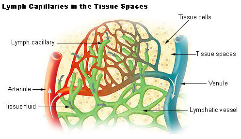
The lymphatic system has three primary functions. First of all, it returns excess interstitial fluid to the blood. Of the fluid that leaves the capillary, about 90 percent is returned. The 10 percent that does not return becomes part of the interstitial fluid that surrounds the tissue cells. Small protein molecules may “leak” through the capillary wall and increase the osmotic pressure of the interstitial fluid. This further inhibits the return of fluid into the capillaries, and fluid tends to accumulate in the tissue spaces. If this continues, blood volume and blood pressure decrease significantly and the volume of tissue fluid increases, which results in edema (swelling). Lymph capillaries pick up the excess interstitial fluid and proteins and return them to the venous blood. After the fluid enters the lymph capillaries, it is called lymph.
The second function of the lymphatic system is the absorption of fats and fat-soluble vitamins from the digestive system and the subsequent transport of these substances to the venous circulation. The mucosa that lines the small intestine is covered with fingerlike projections called villi. There are blood capillaries and special lymph capillaries, called lacteals, in the center of each villus. The blood capillaries absorb most nutrients, but the fats and fat-soluble vitamins are absorbed by the lacteals. The lymph in the lacteals has a milky appearance due to its high fat content and is called chyle.
The third and probably most well known function of the lymphatic system is defense against invading microorganisms and disease. Lymph nodes and other lymphatic organs filter the lymph to remove microorganisms and other foreign particles. Lymphatic organs contain lymphocytes that destroy invading organisms.
Components of the Lymphatic System
The lymphatic system consists of a fluid (lymph), vessels that transport the lymph, and organs that contain lymphoid tissue.
Lymph
Lymph is a fluid similar in composition to blood plasma. It is derived from blood plasma as fluids pass through capillary walls at the arterial end. As the interstitial fluid begins to accumulate, it is picked up and removed by tiny lymphatic vessels and returned to the blood. As soon as the interstitial fluid enters the lymph capillaries, it is called lymph. Returning the fluid to the blood prevents edema and helps to maintain normal blood volume and pressure.
Lymphatic Vessels
Lymphatic vessels, unlike blood vessels, only carry fluid away from the tissues. The smallest lymphatic vessels are the lymph capillaries, which begin in the tissue spaces as blind-ended sacs. Lymph capillaries are found in all regions of the body except the bone marrow, central nervous system, and tissues, such as the epidermis, that lack blood vessels. The wall of the lymph capillary is composed of endothelium in which the simple squamous cells overlap to form a simple one-way valve. This arrangement permits fluid to enter the capillary but prevents lymph from leaving the vessel.
The microscopic lymph capillaries merge to form lymphatic vessels. Small lymphatic vessels join to form larger tributaries, called lymphatic trunks, which drain large regions. Lymphatic trunks merge until the lymph enters the two lymphatic ducts. The right lymphatic duct drains lymph from the upper right quadrant of the body. The thoracic duct drains all the rest.
Like veins, the lymphatic tributaries have thin walls and have valves to prevent backflow of blood. There is no pump in the lymphatic system like the heart in the cardiovascular system. The pressure gradients to move lymph through the vessels come from the skeletal muscle action, respiratory movement, and contraction of smooth muscle in vessel walls.
Lymphatic Organs
Lymphatic organs are characterized by clusters of lymphocytes and other cells, such as macrophages, enmeshed in a framework of short, branching connective tissue fibers. The lymphocytes originate in the red bone marrow with other types of blood cells and are carried in the blood from the bone marrow to the lymphatic organs. When the body is exposed to microorganisms and other foreign substances, the lymphocytes proliferate within the lymphatic organs and are sent in the blood to the site of the invasion. This is part of the immune response that attempts to destroy the invading agent.
The lymphatic organs include:
Lymph Nodes
Lymph nodes are small bean-shaped structures that are usually less than 2.5 cm in length. They are widely distributed throughout the body along the lymphatic pathways where they filter the lymph before it is returned to the blood. Lymph nodes are not present in the central nervous system. There are three superficial regions on each side of the body where lymph nodes tend to cluster. These areas are the inguinal nodes in the groin, the axillary nodes in the armpit, and the cervical nodes in the neck.
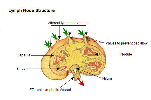
The typical lymph node is surrounded by a connective tissue capsule and divided into compartments called lymph nodules. The lymph nodules are dense masses of lymphocytes and macrophages and are separated by spaces called lymph sinuses. The afferent lymphatics enter the node at different parts of its periphery, which carry lymph into the node; entering the node on the convex side. The lymph moves through the lymph sinuses and enters an efferent lymphatic vessel, which, located at an indented region called the hilum, carries the lymph away from the node.
Tonsils
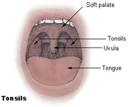
Tonsils are clusters of lymphatic tissue just under the mucous membranes that line the nose, mouth, and throat (pharynx). There are three groups of tonsils. The pharyngeal tonsils are located near the opening of the nasal cavity into the pharynx. When these tonsils become enlarged they may interfere with breathing and are called adenoids. The palatine tonsils are the ones that are located near the opening of the oral cavity into the pharynx. Lingual tonsils are located on the posterior surface of the tongue, which also places them near the opening of the oral cavity into the pharynx. Lymphocytes and macrophages in the tonsils provide protection against harmful substances and pathogens that may enter the body through the nose or mouth.
Spleen
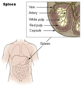
The spleen is located in the upper left abdominal cavity, just beneath the diaphragm, and posterior to the stomach. It is similar to a lymph node in shape and structure but it is much larger. The spleen is the largest lymphatic organ in the body. Surrounded by a connective tissue capsule, which extends inward to divide the organ into lobules, the spleen consists of two types of tissue called white pulp and red pulp. The white pulp is lymphatic tissue consisting mainly of lymphocytes around arteries. The red pulp consists of venous sinuses filled with blood and cords of lymphatic cells, such as lymphocytes and macrophages. Blood enters the spleen through the splenic artery, moves through the sinuses where it is filtered, then leaves through the splenic vein.
The spleen filters blood in much the way that the lymph nodes filter lymph. Lymphocytes in the spleen react to pathogens in the blood and attempt to destroy them. Macrophages then engulf the resulting debris, the damaged cells, and the other large particles. The spleen, along with the liver, removes old and damaged erythrocytes from the circulating blood. Like other lymphatic tissue, it produces lymphocytes, especially in response to invading pathogens. The sinuses in the spleen are a reservoir for blood. In emergencies such as hemorrhage, smooth muscle in the vessel walls and in the capsule of the spleen contracts. This squeezes the blood out of the spleen into the general circulation.
Thymus
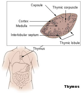
The thymus is a soft organ with two lobes that is located anterior to the ascending aorta and posterior to the sternum. It is relatively large in infants and children but after puberty it begins to decrease in size so that in older adults it is quite small.
The primary function of the thymus is the processing and maturation of special lymphocytes called T-lymphocytes or T-cells. While in the thymus, the lymphocytes do not respond to pathogens and foreign agents. After the lymphocytes have matured, they enter the blood and go to other lymphatic organs where they help provide defense against disease. The thymus also produces a hormone, thymosin, which stimulates the maturation of lymphocytes in other lymphatic organs.
Review: Introduction to the Lymphatic System
- The lymphatic system returns excess interstitial fluid to the blood, absorbs fats and fat-soluble vitamins, and provides defense against disease.
- Lymph is the fluid in the lymphatic vessels. It is picked up from the interstitial fluid and returned to the blood plasma.
- Lymphatic vessels carry fluid away from the tissues.
- The right lymphatic duct drains lymph from the upper right quadrant of the body and the thoracic duct drains all the rest.
- Pressure gradients that move fluid through the lymphatic vessels come from the skeletal muscle action, respiratory movements, and contraction of smooth muscle in vessel walls.
- Lymph enters a lymph node through afferent vessels, filters through the sinuses, and leaves through efferent vessels.
- Tonsils are clusters of lymphatic tissue associated with openings into the pharynx and provide protection against pathogens that may enter through the nose and mouth.
- The spleen is a lymph organ that filters blood and also acts as a reservoir for blood.
- The thymus is large in the infant and atrophies after puberty.